


Osteoarthritis of the hip joint (ATS) is a slow, destructive disease. Irreversible changes in the structure and properties of hyaline cartilage occur during the development of the disease due to several reasons, which leads to an increase in the pressure on the joint surfaces and their deformation or fusion. Given that mechanical overload is considered one of the main causes of the development of the disease, the articulation of the hip joint is often affected by arthrosis.
Characteristics of the anatomical structure of the hip joint
The hip joint (TC) is the meeting point between the pelvis and the femur. This articulation allows lowering and spreading of the lower limbs, raising and pulling the legs to the body, and walking movements. From birth and throughout life, a person carries a large load on the hip joint.
From the side of the pelvic bone, the "acetabular" cavity is involved in the articulation, from the side of the femur in its epiphysis. Along the edges of the acetabulum, there is a collagenous lip that acts as a kind of seal that holds the epiphysis of the femur tightly in the socket. The socket in the center of the acetabulum is covered by a collagen membrane, and the attachment point of the femoral ligament.
The composition of the TS capsule contains tapes:
- femoral-iliac - the strongest ligament that can withstand a load of more than 200 kg and prevents excessive bending of the hip;
- femoral-pubic - responsible for abduction and reduction of the thigh, thereby limiting its circular movements;
- femoral ischialis - protects the vehicle from concussion, reduces the load during walking and running;
- circular (loop) - prevents dislocations and keeps the head of the femur in the pelvic cavity and is the basis of the joint bag.
Several muscle groups and tendons enable the vehicle to move around three axes:
- Longitudinal (vertical).
- Transverse (horizontal, frontal).
- Sagittal (front-back).
Joint arthrosis can also occur in a healthy joint and can be a continuation of existing diseases of the musculoskeletal system.
What is this disease?
Hyaline cartilage performs shock-absorbing and protective functions against damage to the surfaces of the joint. ATS is a disease in which the structure of collagen cartilage changes during its development, which later leads to their fragmentation and destruction. Cartilage fragments, if they enter the joint cavity, can cause an inflammatory process. On bare surfaces, bone tissue undergoes changes caused by friction and increased pressure. The remaining cartilaginous tissue along the edges of the epiphyses grows compensatory with the subsequent ossification, which causes ankylosis (immobility of the bone joint). In the later stages, in the absence of adequate therapy, the patient completely loses the ability to move and becomes disabled. Destructive processes are triggered by various causes.
There are the following types of arthrosis of the hip joint:
- Primary. Its etiology is not fully understood. Idiopathic (primary) arthrosis develops in a previously healthy joint. It most often develops in the elderly.
- Secondary. This is caused by previous diseases of the joint apparatus, congenital malformations, and changes in the work and systems of vital human organs.
The disease develops in one joint or affects both at the same time.
Causes of the disease
Among the causes contributing to the appearance and progression of the disease, the following are identified:
- Hereditary genetic predisposition to the development of the disease.
- Bone joint injuries (dislocations, fractures, sprains and tendons).
- Unbearable systematic force and physical activity.
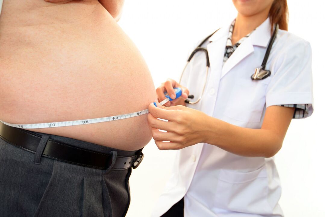
- Overweight.
- Dysfunctions of the endocrine system (diabetes, psoriasis).
- Congenital pathologies of the structure and development of the musculoskeletal system.
- Professional characteristics of the work activity.
- Poor local circulation.
- Previous illnesses caused by pathogenic flora.
- Legg-Calve-Perthes disease.
- Metabolic disorders (gout).
- Physical inactivity.
- Immune diseases.
These reasons may not always cause ATS. Most often, activation of pathological processes can be provoked by:
- increased stress and physical activity;
- constant overtime;
- hypothermia of the vehicle or the body as a whole;
- sudden lifting of heavy objects;
- hormonal imbalance;
- radiation exposure.
Symptoms of the disease
The symptomatic manifestations of ATS are similar to the manifestations of arthrosis of other joints.
The main characteristic symptoms of the disease are as follows:
- Stiffness in the morning or after prolonged immobility.
- Reduced range of motion, change in gait.
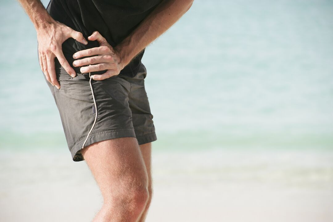
- Pain that is first caused by mechanical or physical stress and then constant.
- Manifestation of crackling, crunching and clicking during sudden movements.
- Marked lameness in the affected limb.
- Occurrence of contractures (restriction of passive movements).
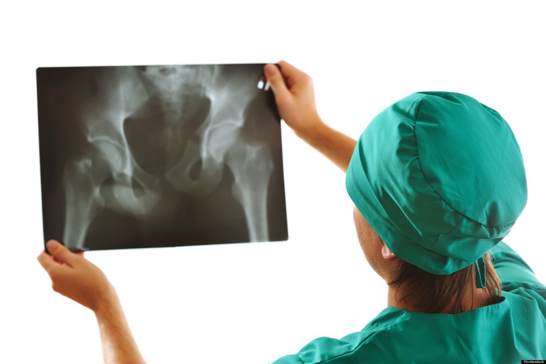
- Narrowing or closing of the joint space (X-ray sign).
The severity of the signs of arthrosis of the hip joint depends on the degree of development of the disease and the reactive capabilities of the patient's body.
Stages of coxarthrosis
Depending on the clinical manifestations, 4 stages of arthrosis of the hip joint can be distinguished:
- Arthrosis of the 1st degree of the hip joint has no pronounced pain and other manifestations. The stage is difficult to diagnose, the disease can be detected by biochemical examination of the hyaline cartilage tissue and determination of the insufficient amount of glycosaminoglycans. The patient feels pain in the joint and rarely feels pain at the beginning of physical activity.
- Second-degree arthrosis of the hip joint is characterized by a change in the density and flexibility of the cartilage. Cracks and fractures appear. Depreciation functions decrease. The pain intensifies, radiates to the lumbar region, and the diluting and reducing movements of the affected limb are limited.
- In the third degree, the layering of cartilage occurs with greater intensity. Joint surfaces experience excessive pressure, ischemia foci develop. Cartilage grows along the edge of the epiphyses. The sensation of pain in the area of the damaged bone connection does not depend on the state of activity and rest. With any movement, the joint "creaks" and "crunches". The range of motion is reduced on all axes.
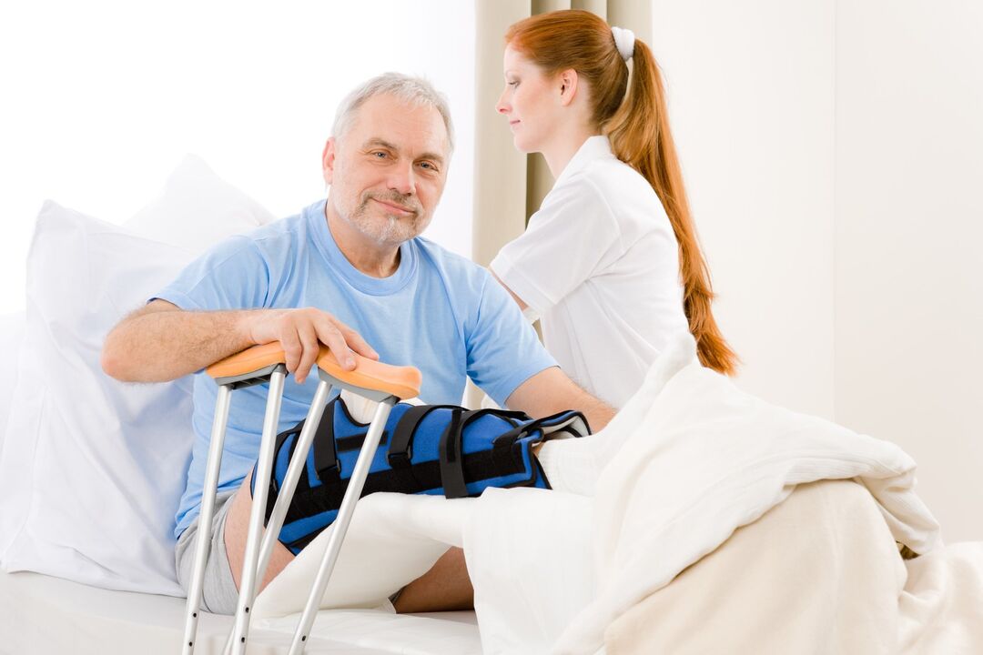
- The fourth degree is characterized by exposure of the surface of the joint components, with the formation of ulcers and depressions. The articular head of the femur is poorly fixed in the acetabulum, which leads to a violation of the comparison and separation of the articular surfaces. During this period, the patient feels unbearable pain, which is caused by the narrowing and sometimes closing of the joint lumen, as well as the compression of bundles of nerve fibers and blood vessels. Movement is limited, sometimes completely.
The classification of pathological changes caused by ATS is necessary to understand the mechanism and characteristics of the disease. Determining the severity of the disease helps to determine the appropriate treatment tactics and disability (in case of severe disease).
Possible consequences
The progression of ATS leads not only to the deformation of the femoral head and pelvic cavity, but also to the development of pathological processes in the functioning of the joint apparatus as a whole.
Pathologies resulting from complications of hip arthrosis:
- synovitis (inflammation of the synovial membrane of the joint);
- aseptic necrosis of the femoral head;
- joint damage (osteonecrosis);
- inflammation of the joint bag with a change in the amount of synovial fluid;
- partial or complete ankylosis (immobility of bone articulation);
- contractures (restriction of mobility and impossibility of bending and extending the limb).
The development of complications of ATS always leads to deterioration of the patient's general condition and quality of life, and loss of movement without assistance.
Diagnostic methods
Diagnosing arthrosis of the hip joint in the initial stage is difficult. Symptomatic manifestations become noticeable only when the epiphyses of bones and nerve fibers are involved in the pathological process.
During the medical examination at the stage of progression, the following are observed:
- visual change in joint contour;
- palpable pain;
- sometimes pastiness of periarticular tissues;
- shortening of the diseased limb.
X-rays play a key role in the diagnosis of ATS. As additional diagnostic methods:
- Ultrasound, magnetic resonance imaging.
- CT scan.
- Puncture of joint lubrication (synovial fluid).
- Diagnosis using an arthroscope (microprobe).
- Clinical and biochemical laboratory examination of urine and blood.
Timely diagnosis improves the prognosis of the treatment and the patient's future life.
How to submit a disability application?
It is impossible to completely cure this disease. To confirm your right to social benefits and assign a disability group, contact your doctor after an examination by narrow specialists.

In the case of hip joint arthrosis, the indication for establishing disability:
- oligoarthrosis (alteration of up to 2 joints) TS 2 degree;
- 2nd degree combined arthrosis of the TS and 3rd degree arthrosis of the knee joint;
- a decrease in the length of the patient's limb by more than 6 cm;
- reactive flow automatic telephone exchange, documented.
It helps to determine the disability group:
- carefully collected anamnesis;
- medical advisory committee (MCC) conclusion;
- results of diagnostic tests;
- passed the Medical and Social Expert Committee (MSEC).
If the expert committee's decision is negative, it can be appealed.
Prevention
Preventive measures are a simple way to avoid developing the disease. Preventive measures include:
- Maintaining an active lifestyle.
- Checking body weight indicators.
- Optimizing nutrition and the way you work and rest.
- Reduced mechanical and physical stress.
- Treatment of diseases of viral and infectious etiology.
- Prevention and prevention of injuries at home and at work.
- Regular preventive examination.
Conclusion
The answer to the frequently asked question: "Is it possible to cure arthrosis of the hip joint? "" The experts give a negative answer. The destroyed cartilage tissue cannot be completely restored, just as the deformation and destruction of the bones in the joint cannot be completely corrected. Do not ignore even minor manifestations of arthrosis of the hip joint, this reduces the chance of preventing the further development of the disease.